Supported by an NIH R01 grant, Biomedical Engineering Professor Dr. Bing Yu combines deep ultraviolet scanning fluorescence microscopy with artificial intelligence to more precisely determine tumor size and shape to improve the accuracy of lumpectomies.
Breast cancer is the most common cancer globally and will affect about one in eight women in the United States over the course of their lifetime. Breast-conserving surgery, also called a lumpectomy, is a common treatment. The goal is to remove only the cancerous tissue while preserving as much of the breast as possible. Although a variety of techniques are available to doctors to use during surgery to determine where a tumor ends and where the healthy tissue starts, existing methods cannot adequately analyze an entire specimen with a high-enough resolution and within the time needed.
As a result, about 20 percent of lumpectomy patients must return for additional surgery to remove various amount of cancerous tissue missed in the original procedure.

“The risk of these additional surgeries can lead to significant emotional, cosmetic and financial burdens for patients and their caregivers,” says Dr. Bing Yu, associate professor in the Marquette University and Medical College of Wisconsin Joint Department of Biomedical Engineering.
Yu and his co-investigators — composed of Dr. Taly Gilat-Schmidt, professor of MU-MCW biomedical engineering, Dr. Dong Hye Ye, assistant professor of computer science at Georgia State University, as well as clinicians Drs. Tina Yen and Julie Jorns from the Medical College of Wisconsin — received a $1.54 million R01 research grant from the National Institutes of Health to develop a novel imaging tool that can more accurately and quickly detect tumor margins during breast conserving surgeries. The technology has the potential to decrease the need for a second surgery as well as emotional stress and costs that may come with it.
About 20 percent of lumpectomy patients must return for additional surgery to remove various amount of cancerous tissue missed in the original procedure… The technology has the potential to decrease the need for a second surgery as well as emotional stress and costs that may come with it.
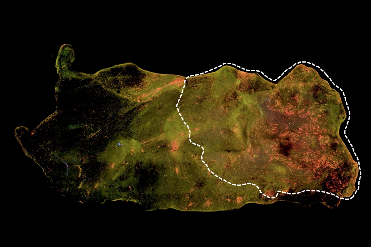
The proposed instrument combines an imaging technology known as deep ultraviolet scanning fluorescence microscopy with the computing capabilities of “deep learning” algorithms. Scientists will first train the computer algorithms, which are connected to the sensors in the microscope, to recognize tissue known to have cancerous cells. The microscope can recognize the cancer because the tissue is stained with special dyes that cause cells and their nuclei to fluoresce under ultraviolet light.
“Our preliminary images demonstrate excellent visual contrasts between breast cancer cells and normal tissues,” says Yu.
According to Yu, the research team published studies showing that the technology can achieve an accuracy of between 93 percent and 96.6 percent in differentiating cancer cells from normal tissues.
Once the algorithms are fully trained in sufficient breast tissue samples, the system can be used to scan new lumpectomy specimens during surgery. The goal is to examine freshly excised tissue specimens and determine whether there is cancer in under five minutes.
Yu sees the final device being compact and easy–to-use, particularly in community hospitals — as opposed to academic hospitals — where the majority of women with breast cancer receive care.
“It should be portable that can be easily moved from room to room in a lab cart or on its own wheels,” he says.
He thinks the technology could be available after four or five more years of research and another few years to go through approval from the Food and Drug Administration.