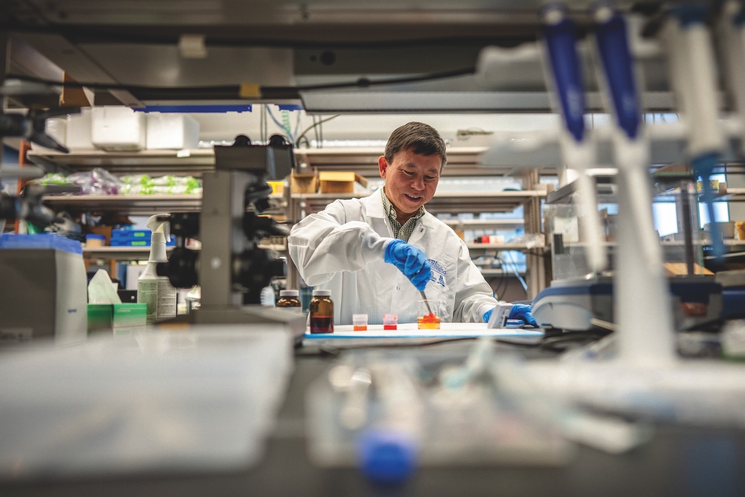
Shortly after joining the Marquette University and Medical College of Wisconsin Joint Department of Biomedical Engineering in 2017, Dr. Bing Yu discovered a breakthrough technology to help solve a thorny clinical problem he’d been working on for years. He also found expert Marquette and MCW colleagues to help him harness that technology.
In Yu’s sights were the many cases of breast-conserving lumpectomy surgeries that leave behind traces of tumors in patients’ bodies. Since these cases are typically caught in post-surgical pathology reports, about 20 percent of lumpectomy patients return for follow-up surgeries to remove the remaining cancer. “These additional surgeries can lead to significant emotional, cosmetic and financial burdens for patients and their caregivers,” says Yu, associate professor of biomedical engineering.

At previous stops in his career, Yu had made intriguing but unfulfilling progress on methods to quickly scan tumor margins of lumpectomy specimens in operating rooms, allowing medical teams to remove tumor remains immediately. His best effort, which obtained simultaneous spectroscopy readings using 49 fiber-optic pinpoints, successfully surveyed large tissue areas quickly, but spacing between data points left gaps where cancer could lurk.
Harnessing a discovery
Then in 2017, a webinar introduced Yu to that breakthrough technology — an ultraviolet microscope that produces vivid images of tissue treated with fluorescent dyes. He determined that the scans could reveal variations in cell size and structure to enable detection of cancer in margins, with effectively no gap in coverage. But this high resolution was a mixed blessing — yielding a flood of visual information exceeding what could normally be processed during surgeries.
As he sought a machine-learning expert with experience building algorithms that automate and accelerate such massive processing tasks, Dr. Dong Hye Ye joined Marquette’s Electrical and Computer Engineering Department. “He had that expertise,” Yu says. “He came at the right time.”
Since joining forces, Yu, Ye (who has since moved to Georgia State University) and partners from MCW have made great strides with a diagnostic tool that offers real hope for improved outcomes for breast cancer patients. With support from a $1.54 million R01 grant from the National Institutes of Health, the team developed a portable system that can be wheeled in and out of surgical rooms. They have determined the device could achieve 96.5 percent accuracy detecting cancer cells in margins of tissue-bank specimens up to 80 square centimeters in surface area — all in 10 minutes or less.

As they develop a next-generation device to extend those rapid results to larger specimens, the team sees more progress ahead. With team members from MCW — breast surgeon Tina Yen, M.D., and pathologist Julie Jorns, M.D. — Yu is writing a grant application that would green-light a clinical trial to test the device in hospitals. And a Way Klingler Research Fellowship is helping him develop a new use for the deep learning-enabled microscope — identifying the presence, and type, of cancer in fresh biopsy samples, without the need for a traditional pathology report. “Many developing countries don’t have access to pathology,” Yu says. “This tool has the potential to lead doctors to a much-needed diagnosis.”
Illustrations by SHOUT