By Alex Nemec, marketing communications specialist
 College of Health Sciences Dean Dr. William Cullinan instructs a participant during a break between didactic instruction.


")

The College of Health Sciences welcomed 100 participants to the 25th running of its popular annual summer brain dissection course, including clinicians, health professionals, educators, researchers and graduate students.
The course, titled Neuroanatomical Dissection: Human Brain and Spinal Cord, ran Aug. 4-6 and attracted attendees from across North America. It is the only course of its kind in the world.
Focused on deepening an understanding of the three-dimensional organization of the central nervous system, the three-day course saw participants spend 12 hours in the lab performing hands-on dissection techniques guided by Marquette faculty. The course also featured topical breakout sessions on recent advances in the neuroscience field provided by CHS faculty researchers.
“Advancing understanding of the organization of the brain through didactic work complemented by this type of dissection experience is a powerful way to approach a complex topic,” says William E. Cullinan, professor and dean of the College of Health Sciences. “It is also extremely useful in helping present and future health care professionals, educators, and researchers better grasp the intricacies of the brain’s internal structure and organization, which can be critical to their professional success.”
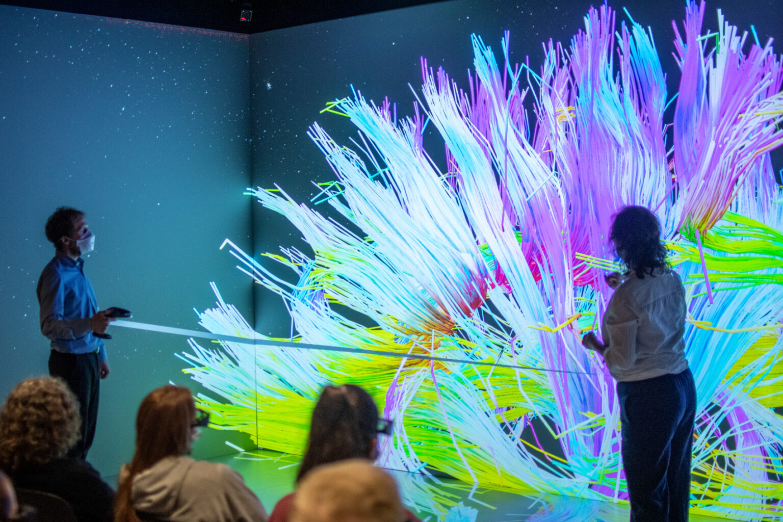
Participants also had the special opportunity to experience a virtual reality simulation of the brain in the Marquette Visualization Lab (MARVL) where Dr. SuJean Choi, professor of biomedical sciences, walked them through a virtual rendering of the brain, including its constituent parts and its white matter fiber pathways.
“The Visualization Lab is always a popular element of the course,” Choi says. “The rendering is vibrant and helps course participants not only better understand the different parts of the brain, but their positions in three-dimensions relative to each other, as well as the functional roles they play in human brain activity.”
To learn more about the neuroanatomical dissection and other continuing education offerings in the College of Health Sciences’ visit here.